Home > Highlighting JAPAN > Highlighting Japan July 2018 > Science & Technology
Highlighting JAPAN
A VR Simulator That Shows Living Hearts at Work
A new viewer that virtually reproduces hearts in 3D and shows their real-time mechanisms with VR technology is already being used as a medical education tool far superior to textbooks and anatomy models, and research on its potential clinical uses is progressing.
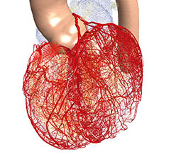
The typical adult heart beats 60 to 80 times per minute, incessantly pumping roughly five liters of blood throughout the body. It is one of the most essential organs in the human body. The heart’s complex structure makes it hard to properly observe the flow of blood and how the organ transmits electronic signals. Heart disease is the second leading cause of death for Japanese people, yet much remains unclear about the illness. That makes research to illuminate the state of the heart vital.
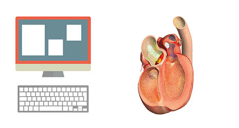
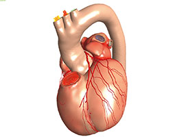
Fujitsu and The University of Tokyo have developed a way to see into the heart in 3D: precise data output by a numerical simulator that shows its real-time movements, blood flow and more. Named Heart Explorer, it went on sale in April 2018 as a medical education tool.
One major feature of Heart Explorer is that the tool doesn’t just construct a 3D model of the heart using a CG creator—it’s an actual “heart” that works thanks to numerical simulation from the K computer. Using mathematical modeling of real heart phenomena, such as the movement of the myocardium, heartbeat, blood output and blood pressure in coronary circulation, Heart Explorer can reproduce the heart’s condition and functioning. It is also possible to reproduce the behavior of a specific patient’s heart using medical images and their diagnostic information.
With the virtual reality display, a 3D model of the beating heart appears that can be turned and rotated as if you were holding it in your hands. It’s possible to zoom in and even view the underside of the heart and the functioning of the aortic valves. In the cross-sectional display, phenomena hard to capture with an MRI or CT scan—such as the state of the myocardium or the blood flow inside the heart—are visible. It’s also possible to overlay real data from an electrocardiogram (ECG) with what has been observed for comparison.
At medical and nursing schools, Heart Explorer is being used as a learning tool to educate students by linking all kinds of data with the structure, condition and functioning of the heart, which is difficult to grasp from textbooks and anatomy models. A few university medical departments, including that of The University of Tokyo, have courses that incorporate Heart Explorer.
Fujitsu’s research into the heart simulator began in 2008 as a collaborative effort with The University of Tokyo. In order to create an app that could solve real-world problems, they began clinical studies using data from heart failure and congenital cardiac disease patients in 2013.
“It was not easy to link up real ECG data with the heart models created from CT scans, MRI and other imaging,” says Masahiro Watanabe, head of Heart Explorer research and development, describing the development process. “We were also constructing extremely complex mathematical models to reproduce the interactions of the myocardium and blood flow, as well as the state of the excitatory signals—the electronic signals that tell the heart to beat.”
Fujitsu is researching and developing clinical uses for Heart Explorer as well.
“Before, heart simulation technology only consisted of tools to explain things to patients who’d already completed treatment,” says Watanabe, “but we also want this research to be useful in diagnosis and treatment. It became possible to find the optimal location for CRT (cardiac resynchronization therapy) electrodes for patients who have pacemakers, and we are considering future uses such as predicting the postoperative status of the heart by performing virtual surgery on models made from real patients’ hearts.”
Clinical use raises the issues of effectiveness, safety and cost. Compared to the mathematical processing of the past that took roughly ten days to calculate five full cardiac cycles, however, today’s simulation and visualization technology can do it in ten hours, and is progressing at light speed. As the value of 3D heart model visualization gains more recognition, research into its clinical applications is expected to continue to evolve.


No article or any part there of may be reproduced without the express permission of the Cabinet Office. Copyright inquiries should be made through this form.
© 2009 Cabinet Office, Government of Japan